艺术
美容与健康
工艺
文化与历史
娱乐
环境
食品与饮料
逆向工程
科学
体育
技术
可穿戴设备
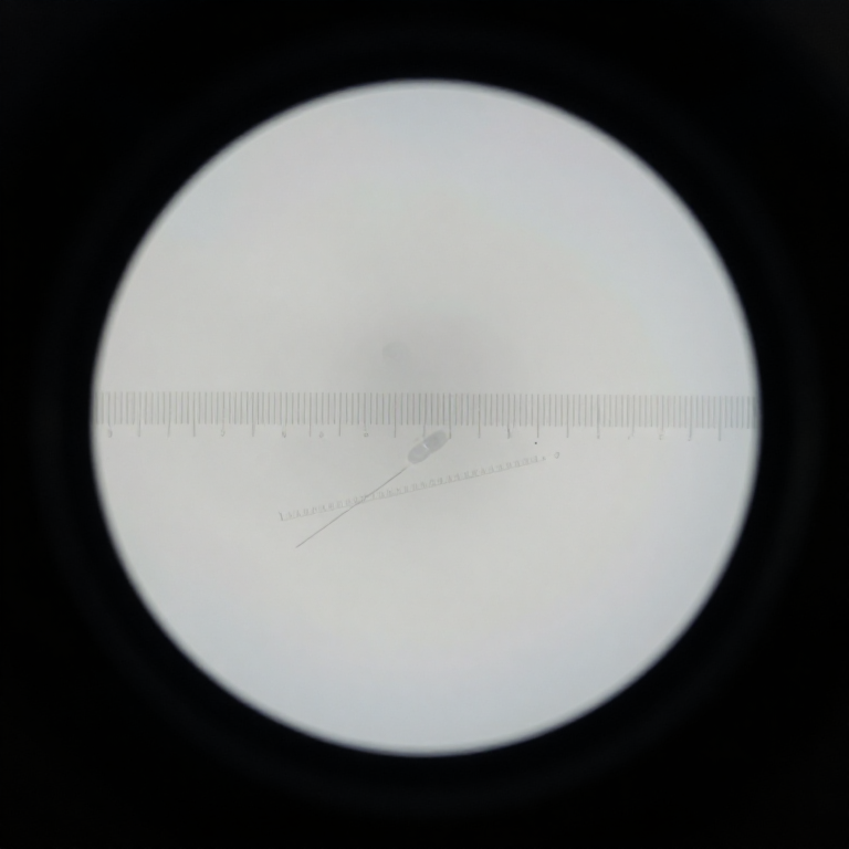


Micrometry: Measuring with a Microscope
Seven rungs of this batch make things visible. This one turns looking into measuring, and it is the rung that is almost always skipped.
A microscope has no inherent scale. The number stamped on the objective is nominal, the tube length is approximate, and 'about the size of a red blood cell' is not a measurement. What you need is an eyepiece graticule — a ruled scale in the eyepiece, which moves with the image — calibrated against a stage micrometer of known spacing.
Do it once per objective and write the numbers on a card taped to the microscope, and every observation you make afterwards carries a dimension. That is the difference between a hobby and an instrument, and it is the same argument the turbidity tube and the reference library made in the two batches before this one.
初学者
3 hours
说明
1
1
Fit the eyepiece graticule
Fit the eyepiece graticule
Unscrew the eyepiece's top lens, drop the etched glass disc onto the internal shoulder with the ruled side facing the field lens, and reassemble.
It must sit at the eyepiece's field stop — the plane where the intermediate image forms — or it will never be in focus at the same time as the specimen. If the scale and the specimen cannot both be sharp, the disc is in the wrong plane, not the wrong eyepiece. Focus the scale itself by turning the eyepiece's top lens before you focus the microscope.
此步骤所需材料:
 刻度分划板(蚀刻玻璃)1 个
所需工具:
 显微镜
显微镜2
2
Make a stage micrometer if you cannot buy one
Make a stage micrometer if you cannot buy one
A stage micrometer is a slide with a finely ruled scale of known spacing. If you do not have one, a diffraction grating of stated line spacing, or a printed scale photographed and reduced by a known factor, will serve — but measure it, do not assume it.
Whatever you use becomes the origin of every dimension you will ever quote. An error here propagates into all of them silently and identically, which is the worst kind: it never shows up as inconsistency. This is the same argument as the home-made turbidity standard, with the same honest caveat.
此步骤所需材料:
 载玻片及盖玻片5 个
所需工具:
 直尺
直尺3
3
Calibrate each objective separately
Calibrate each objective separately
Put the stage micrometer on, focus, and line the two scales up side by side. Count how many EYEPIECE divisions span a known number of STAGE divisions, using as long a span as the field allows.
Use the longest span you can, not the shortest: reading 80 eyepiece divisions against 10 stage divisions divides your reading error by eight. Repeat for every objective — the calibration is different for each, and it changes again if you swap the eyepiece.
所需工具:
显微镜4
4
Write the card and tape it to the microscope
Write the card and tape it to the microscope
One line per objective: objective, eyepiece, micrometres per eyepiece division. Tape it to the base.
A calibration you have to look up is a calibration you will not use, and a measurement made with a half-remembered factor is worse than none. The card also records WHICH eyepiece — swap in a different one and every number on it is wrong, which is the commonest way a careful calibration quietly stops being true.
此步骤所需材料:
 卡纸2 个
卡纸2 个5
5
Measure a real population, not one cell
Measure a real population, not one cell
Measure thirty or more of whatever you are interested in — yeast cells, pollen grains, fibre diameters — and record every value, not a typical one.
Biological things vary, often by a factor of two, so a single measurement tells you almost nothing and 'about 5 micrometres' hides whether the population is tight or spread. The spread is frequently the interesting part: it is how you tell one species from another, or a healthy culture from a stressed one.
此步骤所需材料:
 面包酵母10 克载玻片及盖玻片10 个
所需工具:
显微镜6
6
Calibration, error and how many to count
Calibration, error and how many to count
正在加载 Jupyter 笔记本…
7
7
Quote the measurement the way it can be checked
Quote the measurement the way it can be checked
Write: the value, the spread, how many you counted, the objective and eyepiece, and the calibration factor you used.
'Yeast cells 5.2 ± 1.3 µm, n = 34, 40× with 10× graticule, 0.303 µm per division' is a statement another maker can repeat and disagree with. '5 microns' is not. This is the same standard the reference-library rung set for identification, and it is what makes an observation worth feeding to MOS rather than just worth looking at.
相关蓝图
这些蓝图共享知识——技术、材料或原理




CC0 公共领域
此蓝图以 CC0 协议发布。你可以自由复制、修改、分发和使用此作品,无需征得许可。
通过购买蓝图中的产品支持创客,他们将获得 创客佣金 (由供应商设定),或创建此蓝图的新版本并将其作为连接包含在你自己的蓝图中以分享收入。