

Osmosis and Plasmolysis
Put a strip of potato in salty water and it goes limp. Put an identical strip in plain water and it goes stiff. Nothing was added to the potato and nothing was taken out by hand — water moved on its own, and it moved in opposite directions in the two beakers.
The mechanism is osmosis: water crossing a membrane that lets water through but not the dissolved substance, always in the direction that dilutes the more concentrated side. It is not a pump and it costs the cell nothing. It is simply the statistically inevitable result of water molecules moving at random through a barrier that is one-way for salt.
A plant cell has a second component that changes the outcome completely: a rigid cell wall outside the flexible membrane. Water entering makes the cell swell against the wall and stop — the cell becomes turgid, and turgor is what holds a lettuce leaf up. Water leaving lets the membrane peel away from the wall, and that visible separation is plasmolysis.
The experiment is one of the few where you can measure the effect quantitatively on a kitchen scale and watch it happen under a microscope. Do both, on the same afternoon, and osmosis stops being a definition to memorise.
手順
Cut identical potato strips
Cut identical potato strips
Cut six strips of potato as close to identical as you can — same length, same cross-section. Blot them dry and weigh each to 0.01 g, recording individually.
Number them.
Blotting matters more than it sounds. Surface water is several per cent of a small strip's mass, and inconsistent blotting will swamp the effect you are trying to measure.
このステップの材料:
 じゃがいも2 個
必要な工具:
 デジタルスケール
 デジタルノギス 6インチ
Build a concentration series
Build a concentration series
Make up salt solutions at roughly 0, 1, 2, 5, 10 and 20 % by mass. Put one strip in each, fully submerged, and leave for an hour or more.
Keep them all at the same temperature.
A single salty beaker only shows that something happens. A series shows you where the direction of water movement reverses, and that crossing point is the actual measurement.
このステップの材料:
 細粒の海塩1 パック
Reweigh and find the crossing point
Reweigh and find the crossing point
Blot each strip exactly as before and reweigh. Compute the percentage change in mass and plot it against solution concentration.
Expect a line crossing zero somewhere in the low percentages.
The concentration at which the strip neither gains nor loses mass is the concentration inside the potato's own cells. You have measured the internal solute concentration of a living tissue without opening a single cell.
必要な工具:
 方眼紙
方眼紙Mount onion epidermis
Mount onion epidermis
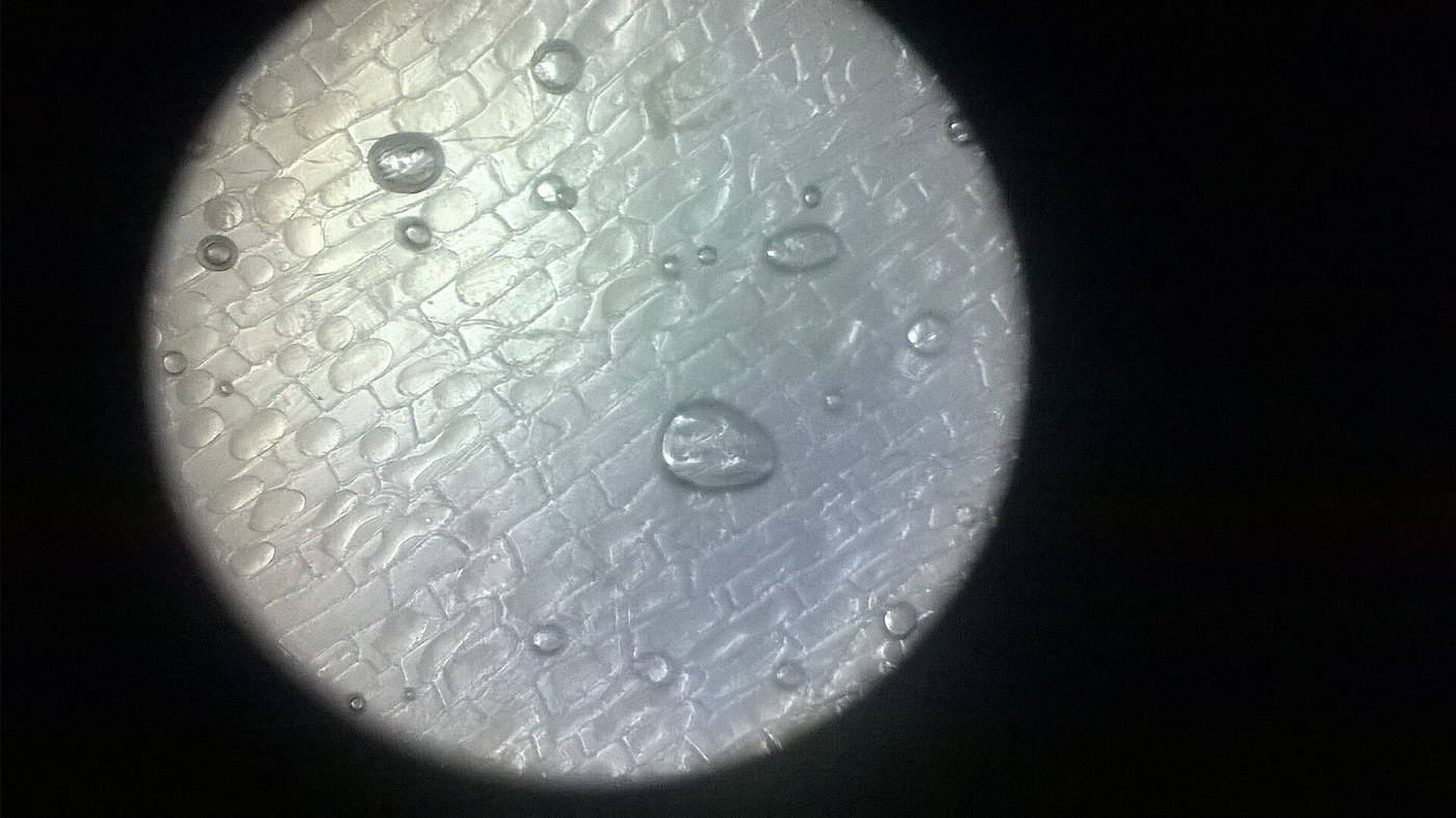
Snap a piece of onion and peel the thin, transparent skin from the inner curve of a scale. Lay a small piece flat on a slide in a drop of water, without folding it, and add a cover.
Focus at the highest magnification you have.
Expect a tidy grid of brick-shaped cells with a visible wall and a nucleus. Onion epidermis is one cell thick, which is exactly why it is the classic specimen.
このステップの材料:
 タマネギ1 個
必要な工具:
 スライドガラス
 顕微鏡
顕微鏡 先の尖ったピンセット
Watch plasmolysis happen live
Watch plasmolysis happen live
Without moving the slide, draw strong salt solution under the cover slip from one edge with a tissue at the other, and keep watching.
Expect the cell contents to shrink away from the wall over a minute or two, leaving a clear gap.
You are seeing the membrane, indirectly. It is too thin to resolve, but its position is now visible because it has separated from the wall it was pressed against.
Reverse it, and find the limit
Reverse it, and find the limit
Flush the same slide with plain water and keep watching.
Expect partial recovery — the cells re-expand.
Now plasmolyse a fresh sample hard and for much longer, then flush.
Expect this one to recover poorly or not at all.
Mild plasmolysis is reversible and severe plasmolysis kills the cell. That is the whole reason salted ground stays barren and why over-fertilising scorches a plant.
Compendium — osmosis, turgor and where the model ends
Compendium — osmosis, turgor and where the model ends
Why water moves at all. Water crosses a semipermeable membrane down its own concentration gradient — from where water is more abundant to where it is less. Adding solute reduces the effective concentration of water, so water moves towards the solute. The formal quantity is water potential, which combines the solute effect with any physical pressure; water always moves from higher to lower water potential. No energy is spent by the cell, which is why osmosis works just as well in dead tissue as living tissue — an important check on the common belief that it is something the cell does.
Turgor is a structural material. An animal cell in pure water swells and bursts. A plant cell cannot, because the cellulose wall resists: internal pressure rises until it balances the osmotic pull, and the cell stops taking water. That pressure is turgor, and it is genuinely load-bearing — a non-woody plant stands up because millions of pressurised cells are pushing outwards against their walls, exactly like an inflatable structure. Wilting is turgor loss, and it is reversible until it is not.
The same physics, in four unrelated places. Root hairs take up soil water osmotically. Kidney tubules and dialysis both work on membrane selectivity. Preserving food with salt or sugar works by making the surroundings osmotically hostile to bacteria and fungi. And a slug shrivels on salt for precisely the reason your onion cells do. The reason osmosis is taught early is not that it is simple but that it is everywhere.
Honest limits of the potato experiment. The strips lose and gain solute as well as water, so the crossing point is an estimate rather than an exact internal concentration. Potato varies with variety, age and storage — old stored potatoes give a different answer from fresh ones. Cutting damages the outermost cells, which leak. And the cell wall is fully permeable to salt, so the barrier you are studying is the membrane just inside it, not the wall — a distinction that a first look at plasmolysis makes unusually clear.
Practical notes. Iodine or a food dye makes onion nuclei far easier to see, and a little dye in the salt solution makes the plasmolysed gap obvious. Work with clean hands, do not eat the salted samples, and use the lowest light that gives a clear image — strong microscope illumination heats a thin specimen and will kill the cells you are trying to watch recover.
接続ブループリントの材料
CC0 パブリックドメイン
このブループリントはCC0で公開されています。許可を求めずに、自由にコピー、修正、配布、あらゆる目的で使用できます。
メイカーを応援するには、ブループリント経由で製品を購入してください。メイカーには メイカーコミッション がベンダーにより設定されています。または、このブループリントの新しいイテレーションを作成し、自分のブループリントにコネクションとして含めて収益を共有できます。