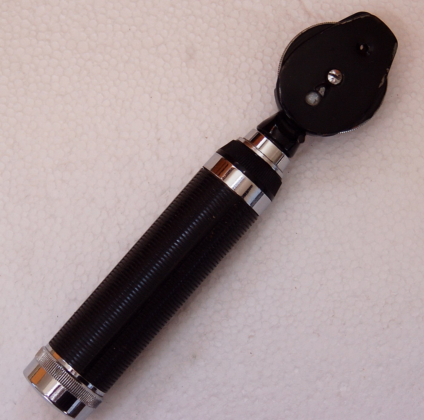


Ophthalmoscope
Зааварчилгаа
Show why the pupil looks black
Show why the pupil looks black
Model the eye as a box with a lens and a small aperture.
- Make a light-tight box with a small hole and a lens behind it.
- Line the back with pale card to stand in for the retina.
- Shine a torch at the hole from beside your head and try to see the back wall.
Энэ алхмын материал:
 Гүдгэр линз1 ширхэг
Put the observer in the beam
Put the observer in the beam
Helmholtz's insight: look along the illuminating axis, not beside it.
- Set a partly reflecting surface — a thin glass plate — at 45 degrees between your eye and the model.
- Shine the torch in from the side, so the plate reflects it into the aperture.
- Look THROUGH the plate along the same axis.
Энэ алхмын материал:
 Хавтгай толь1 ширхэг
 Акрилын хавтан1 хуудас
Add the focusing lenses
Add the focusing lenses
Patients are short-sighted, long-sighted or neither, and so are examiners.
- Add a holder that can swing different lenses into the viewing path.
- Change the model eye's internal length to simulate a refractive error.
- Select the lens that brings the back wall into focus.
Understand what the red reflex means
Understand what the red reflex means
Do NOT examine anyone's eyes with a homemade instrument — but know what clinicians look for.
- A healthy retina returns a uniform orange-red glow, the red reflex — this is the same effect as red-eye in flash photography.
- An absent or white reflex is a serious sign, and in a child can indicate retinoblastoma.
- Clinicians examine the optic disc, the vessels and the macula in turn.
History and context
History and context
Hermann von Helmholtz built his Augenspiegel in 1851, at 30, while teaching physiology at Königsberg — reportedly while preparing a lecture demonstration on why the pupil appears black. He was one of the most wide-ranging scientists of the century: the conservation of energy, the theory of hearing, colour vision, nerve conduction speed and the physiology of the eye all carry his work.
He was not quite first, and he said so. Charles Babbage — the computing pioneer — had built a similar instrument around 1847 and shown it to a physician who apparently did not grasp its value; it went nowhere. Priority in instruments often belongs to whoever puts the device into the hands of people with a use for it, and Helmholtz published into a medical community that immediately understood what it had.
It created a speciality. Before 1851, ophthalmology inferred internal disease from external signs and the patient's account. Afterwards it could observe pathology directly in a living person. That is a rare kind of instrument: not a better measurement of something already visible, but a first sight of something previously hidden.
Where it went: the fundus camera photographs what the ophthalmoscope shows, optical coherence tomography images the retina in cross-section micrometre by micrometre, and automated screening now examines retinal photographs for diabetic damage at population scale. All of them look through the pupil along the illumination axis — the geometry Helmholtz worked out.
Материал
3- 1 ширхэгТүр орлуулагч
- 1 ширхэгТүр орлуулагч
- 1 хуудасТүр орлуулагч
Холбоотой загварууд
Эдгээр загварууд мэдлэг хуваалцдаг — арга техник, материал эсвэл зарчим


CC0 Нийтийн домэйн
Энэ загвар CC0 дор гаргагдсан. Та зөвшөөрөл авахгүйгээр хуулах, өөрчлөх, түгээх, ашиглах боломжтой.
Загвараар дамжуулан бүтээгдэхүүн худалдаж авч Бүтээгчийг дэмжээрэй Бүтээгчийн шимтгэл Борлуулагчаар тогтоосон, эсвэл энэ загварын шинэ хувилбар үүсгэж орлогоо хуваахын тулд өөрийн загварт холбоос болгон оруулна уу.