ARTE
BELEZA E BEM-ESTAR
ARTESANATO
CULTURA E HISTÓRIA
ENTRETENIMENTO
MEIO AMBIENTE
COMIDA E BEBIDAS
ENGENHARIA REVERSA
CIÊNCIAS
ESPORTES
TECNOLOGIA
TECNOLOGIA VESTÍVEL

Firestone Pulse-Echo Reflectoscope
Everything else in this batch shines something THROUGH the patient and catches what comes out the far side. This one stands on one side and listens for the echo, and it arrived in medicine sideways - from a man trying to find cracks inside metal castings.
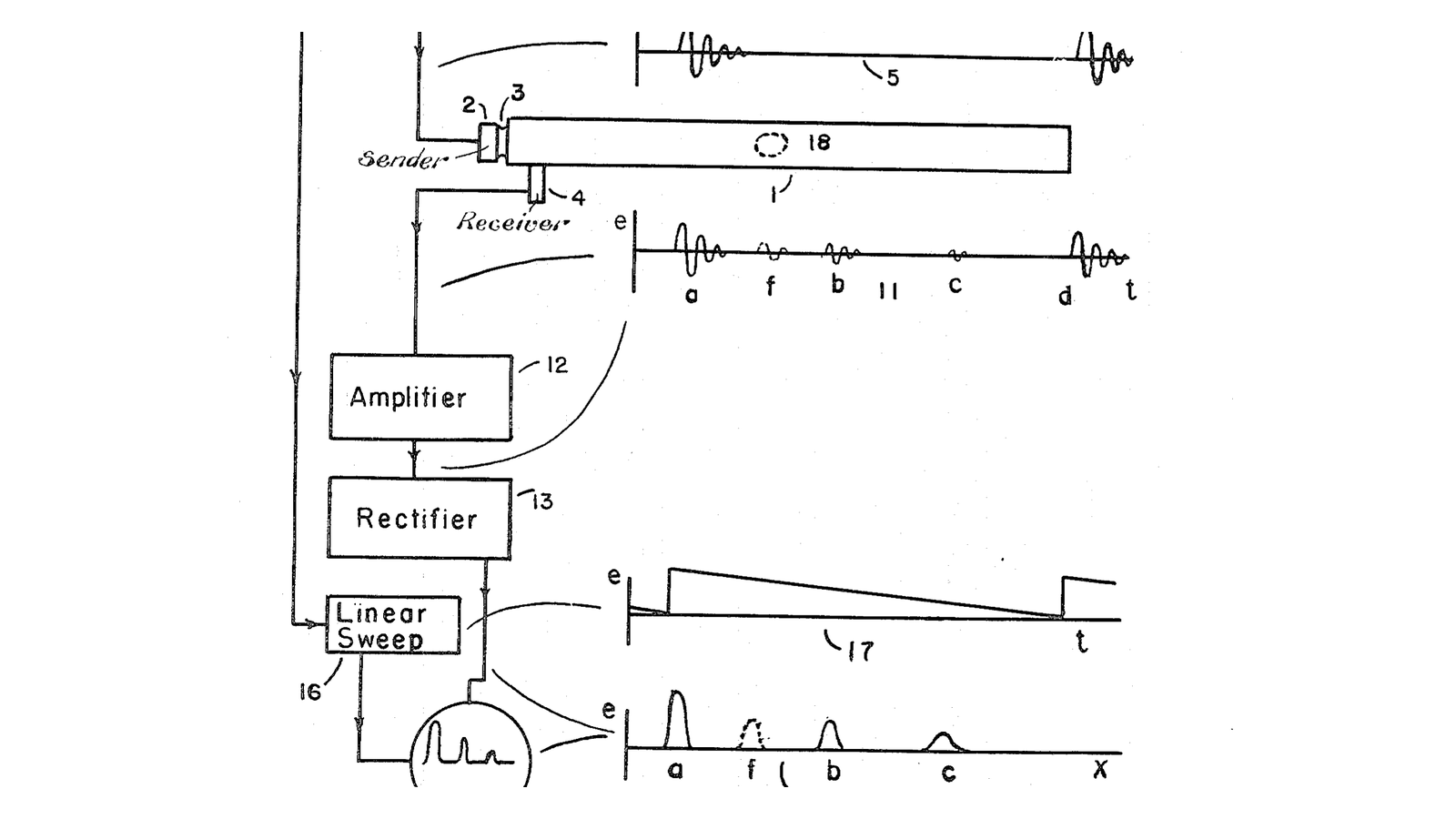
**US 2,280,226**, "Flaw detecting device and measuring instrument", Floyd A. Firestone, filed 27. May 1940 and granted 21. April 1942. Its first claim is a complete description of every ultrasound machine ever built: *"means for transmitting a supersonic wave train into said part, and means for determining the time interval between the sending out of said wave train and the arrival of said wave train at some point of the part"*. Send a pulse. Time the echo. That is all of it.
The drawing on this blueprint is his Figure 1 and it is worth studying, because it is an A-scan - the display every ultrasound machine still produces internally - drawn in 1940. A pulse oscillator gates a high-frequency oscillator; the burst goes into the part; echoes come back from the front face, from the flaw, and from the back wall; they are amplified, rectified, and painted against a linear sweep so that **horizontal distance on the screen is depth into the object.**
It took a decade and a half for the medical use to follow. John Wild and John Reid in the United States were working on tissue echoes by the early 1950s, and Ian Donald in Glasgow brought obstetric scanning into practice in the late 1950s and 60s using industrial flaw detectors of exactly Firestone's type - the machines were already sitting in the shipyards on the Clyde.
**You will build an A-scan and measure with it.** You will find the speed of sound in a material by timing an echo through a known thickness, then use that number to locate something you cannot see. And you will meet the trade that decides every probe ever made: high frequency gives fine detail and cannot get deep; low frequency reaches but cannot resolve.
A sibling worth reading beside this one is the ultrasonic parking sensor already in the catalogue. Same physics, one target, in air, where the speed is 343 m/s and there is no coupling problem. Here the medium is dense, the echoes are many, and getting the sound IN at all is most of the difficulty.
Intermediário
3 hours
Instruções
1
1
Getting the sound in: why coupling is not optional
Getting the sound in: why coupling is not optional
Before any electronics, understand the wall you are up against. A sound wave arriving at a boundary is reflected by an amount set by the mismatch in **acoustic impedance** between the two materials - density times speed of sound.
Tissue against air mismatches by a factor of about four thousand. Run the numbers in the notebook step: the reflection at a tissue-air interface is over 99 per cent. A probe held dry against skin does not give a weak image, it gives no image, because essentially none of the sound ever enters.
The fix is a coupling layer with an impedance close to the target - water, or a gel that is mostly water. That is all ultrasound gel is, and it is doing the single most important job in the system.
Set up a shallow tank of water as your test bed. Working through water removes the coupling problem entirely, lets you move a target to a measured distance, and is how every ultrasound test object is characterised.
Materiais para este passo:
 Recipiente de plástico (hermético)1 peça
 Água destilada2000 ml
 Gel de ecografia1 peça
Ferramentas necessárias:
 Régua
Régua Paquímetro digital de 6 polegadas
2
2
Pulse it and see the echo
Pulse it and see the echo
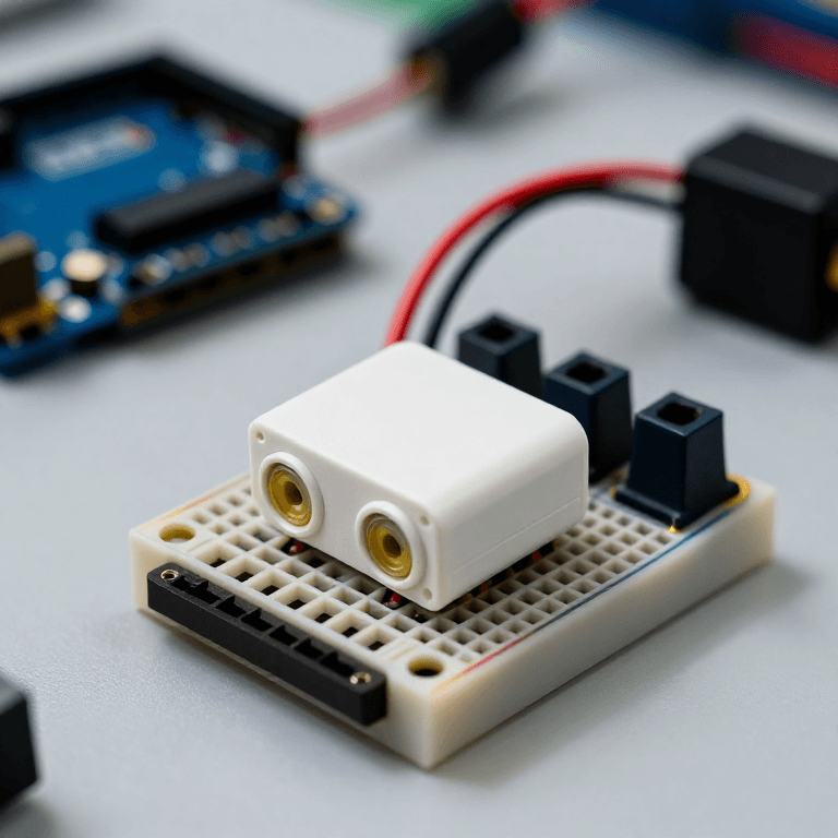
An HC-SR04 module is the cheapest source of a matched pair of 40 kHz transducers. Use the two cans on it and ignore the module's own logic, which reports one distance and hides the echo train you came to see. Drive one with a short burst - five to ten cycles - from a function generator or a microcontroller, then listen on a second transducer or on the same one after the drive stops.
Watch it on an oscilloscope. You will see the transmit burst as a large spike, then a period where the transducer is still ringing and useless, then - if everything is right - a smaller echo.
**That ring-down is a real limitation, not a fault of your build.** A transducer that has just been driven hard keeps vibrating, and while it does it cannot hear. Anything closer than the ring-down time is invisible, which sets a minimum depth for every pulse-echo system ever made. Look at Firestone's Figure 1: the first large spike marked *a* is exactly this, and every echo he cares about comes after it.
40 kHz is far below medical frequencies and that is deliberate - the transducers are cheap, the timings are easy to see, and the physics is identical. The notebook step shows what changing frequency would buy and cost.
Materiais para este passo:
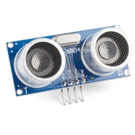 Ultrasonic Sensor - HC-SR041 peça
Ferramentas necessárias:
 Gerador de funções
 Osciloscópio
 Multímetro digital de laboratório
 Cabos de ensaio com pinças jacaré
 Placa de ensaio
3
3
Calibrate: measure the speed of sound in your medium
Calibrate: measure the speed of sound in your medium
Time is only depth if you know the speed, so measure it rather than looking it up.
Put a flat reflector - an acrylic plate - in the tank at a measured distance from the transducer. Read the echo time from the oscilloscope. The sound went there and back, so **speed = 2 × distance ÷ time**.
Do it at five distances and plot time against distance. You should get a straight line through the origin whose slope gives the speed, and using five points rather than one removes the fixed delays in your electronics, which otherwise show up as a constant offset and corrupt a single measurement.
Compare your answer with 1480 m/s for water. Then look at the notebook table and note that every clinical scanner assumes 1540 m/s for ALL soft tissue - fat is slower, muscle faster - so every scanner is slightly wrong everywhere, in a way that is well understood and accepted.
Materiais para este passo:
 Chapa de acrílico1 peça
Ferramentas necessárias:
OsciloscópioRéguaPaquímetro digital de 6 polegadas4
4
Find something you cannot see
Find something you cannot see
Now use it as an instrument rather than a demonstration.
Set up a target you cannot see: a block of gelatine or ballistics gel with an object embedded in it, or simply a closed opaque bottle with a layer of oil floating on water. Both give you an internal interface at an unknown depth.
Couple the transducer to it with gel, pulse, and record every echo time you can see. Convert each to a depth using the speed you measured in step 3. Then open the target and measure the real depths with a ruler.
**Write both columns down side by side.** That comparison - predicted against actual - is the entire claim of the technique, and it is the first time in this batch that you get a NUMBER for something inside an object rather than a picture of it.
If you have a two-axis slide, repeat the measurement at twenty positions across the target and plot depth against position. You have just made a B-scan by hand: a cross-section built out of many A-scans, which is exactly what Wild, Reid and Donald did mechanically.
Materiais para este passo:
 Gelatina em pó200 g
 Óleo alimentar200 ml
 Anilha plana3 peçasGel de ecografia1 peça
Ferramentas necessárias:
OsciloscópioRéguaPaquímetro digital de 6 polegadas5
5
Depth, resolution, and why probes come in sets
Depth, resolution, and why probes come in sets
A carregar o notebook Jupyter…
6
6
When there is no echo
When there is no echo
Three faults account for almost every silent trace, and the first is the reason gel exists.
Flow
Loading...
Ferramentas necessárias:
Osciloscópio7
7
Compendium: from castings to obstetrics
Compendium: from castings to obstetrics
**Why industry got there first.** A steel casting is uniform, has flat parallel faces, does not move, does not mind being clamped in a water bath, and reflects beautifully. A patient is none of those things. Firestone's instrument worked immediately on metal and needed fifteen years of development before it worked on people.
**A-mode, B-mode, M-mode.** A-mode is what you built: amplitude against time, one line, no picture - still used for precise eye measurements before cataract surgery. B-mode turns each echo into a bright dot and sweeps the beam to build a cross-section, which is the familiar image. M-mode plots one line against time to show MOVEMENT, which is how a heart valve is assessed. All three come from the same echo train; only the display changes.
**Why the picture is grainy.** That texture is speckle, and it is not noise in the usual sense - it is the interference pattern of echoes from structures far smaller than the wavelength. Averaging more frames does not remove it, because it is reproducible. It also carries information: experienced sonographers read tissue character from the speckle pattern itself.
**What ultrasound uniquely has.** No ionising radiation, real time, portable and cheap. It is the only modality in this batch that can be brought to the patient rather than the patient to it, which is why it is the one that reaches rural clinics and disaster zones - and why a hand-held probe on a phone is a serious instrument rather than a toy.
**What it cannot do.** Air and bone are walls. Lungs, bowel and anything behind a rib are effectively invisible, and no amount of power fixes it - the sound is reflected at the boundary, not absorbed on the way. That is a hard limit set by the impedance table in the notebook, and it is why the chest and the skull are still X-ray and MRI territory.
Materiais
8- Referência
- 2000 mlReferência
- 1 peçaReferência
- Ultrasonic Sensor - HC-SR0410% de comissão1 peçaR$20.48
- 1 peçaReferência
- 200 gReferência
- 200 mlReferência
- 3 peçasReferência
Ferramentas necessárias
7- Referência
- Referência
- Referência
- Referência
- Referência
- Referência
- 1 vendor sell this, none ship to you yetReferência
Total estimado
O que quem fez comprou. Os materiais sem preço obtêm-se onde os comprares.
Blueprints relacionados
Estes blueprints compartilham conhecimento — técnicas, materiais ou princípios


CC0 Domínio Público
Este blueprint é liberado sob CC0. Você é livre para copiar, modificar, distribuir e usar este trabalho para qualquer finalidade, sem pedir permissão.
Apoie o Maker comprando produtos através do Blueprint, onde ele ganha uma Comissão Maker definida pelos vendedores, ou crie uma nova versão deste Blueprint e inclua-o como conexão no seu próprio Blueprint para compartilhar receita.