

Electrospinning — Making Nanofibers with Electric Fields
Electrospinning is a fiber-formation process that uses a high-voltage electric field to draw a jet of polymer solution from a needle tip into ultra-fine fibers — typically 50 to 500 nanometers in diameter, roughly 1,000 times finer than a human hair and 10–100 times finer than conventional microfibers. The phenomenon was first observed by John Francis Cooley (US patent 692,631, 1902) and William James Morton (US patent 705,691, 1902), but it remained a laboratory curiosity for decades. In the 1990s, Darrell Reneker at the University of Akron systematically studied the process physics, and commercial interest exploded as researchers discovered that nanofiber membranes had extraordinary properties for filtration, biomedical scaffolds, and energy applications.
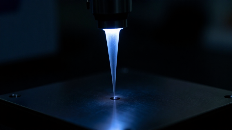
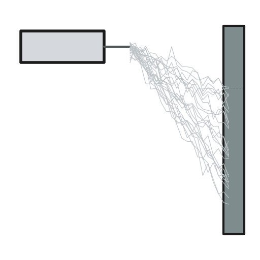
The setup is remarkably simple. A polymer solution (or melt) is loaded into a syringe with a metallic needle. A high voltage (10–30 kV) is applied between the needle tip and a grounded collector plate. The electric field charges the solution surface at the needle tip, forming a conical meniscus called the Taylor cone. When the electrostatic repulsion overcomes the surface tension, a thin jet erupts from the cone tip. As the jet travels toward the collector, it undergoes a chaotic whipping instability that stretches it by a factor of 10,000–100,000, producing a continuous nanofiber that deposits on the collector as a nonwoven membrane.
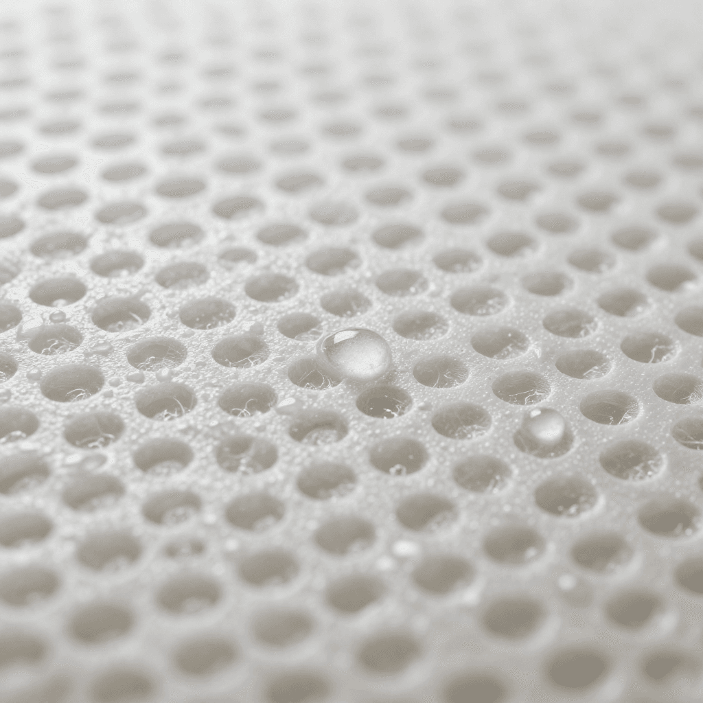
Nanofiber membranes have surface areas of 1–100 square meters per gram — hundreds of times more than conventional fabrics. This extreme surface area, combined with nanoscale pore sizes, makes them ideal for air and liquid filtration (capturing particles that pass through conventional filters), wound dressings (mimicking the extracellular matrix for tissue regeneration), drug delivery systems, battery separators, catalytic supports, and protective clothing. Electrospinning has moved from laboratory to commercial production: companies like Elmarco, Inovenso, and Revolution Fibres operate industrial multi-needle and needleless electrospinning lines producing nanofiber membranes at rates of hundreds of square meters per hour.
안내
Understand the nanoscale
Understand the nanoscale
A nanometer is one billionth of a meter — one millionth of a millimeter. A human hair is approximately 70,000 nanometers in diameter. A standard polyester textile fiber is 10,000–20,000 nanometers. A microfiber is 1,000–10,000 nanometers. An electrospun nanofiber is 50–500 nanometers — approaching the diameter of a single collagen fibril in human skin (50–100 nm). At this scale, the surface-area-to-volume ratio becomes enormous: a single gram of nanofibers with 100 nm diameter has a surface area of approximately 40 square meters. This extreme surface area drives most of the nanofiber's unique functional properties.
Prepare the polymer solution
Prepare the polymer solution
Dissolve a fiber-forming polymer in a volatile solvent to create a spinning solution (spinning dope). Common systems include: polyvinyl alcohol (PVA) in water; polycaprolactone (PCL) in chloroform/methanol for biomedical scaffolds; polyacrylonitrile (PAN) in dimethylformamide (DMF) for filtration; nylon 6 in formic acid; polyurethane in DMF/THF for elastic nanofibers. The polymer concentration (typically 5–20% by weight), molecular weight, and solvent properties determine whether the jet forms fibers, beaded fibers, or droplets. The solution must have enough chain entanglement to sustain a continuous jet — too dilute and it breaks into droplets (electrospray instead of electrospinning).
이 단계의 재료:
 에틸렌글리콜100 ml
필요한 도구:
 유리 증류 플라스크
Set up the electrospinning apparatus
Set up the electrospinning apparatus
The basic electrospinning setup consists of three components: a syringe pump that delivers the polymer solution at a controlled flow rate (0.1–5 mL/hour) through a blunt metallic needle (18–25 gauge); a high-voltage power supply (0–30 kV DC) connected to the needle; and a grounded collector (a flat metal plate, rotating drum, or mandrel) positioned 10–25 cm from the needle tip. The entire setup fits on a laboratory bench. The simplicity is deceptive — the physics of the jet are extraordinarily complex, involving electrostatics, fluid mechanics, and polymer chain dynamics simultaneously.
Form the Taylor cone and initiate the jet
Form the Taylor cone and initiate the jet
Apply voltage to the needle while the syringe pump delivers solution. At the needle tip, the electric field charges the solution surface. The droplet deforms from a hemisphere into a cone — the Taylor cone, named after Sir Geoffrey Taylor, who calculated its theoretical half-angle of 49.3° in 1964. At a critical voltage (typically 8–15 kV for a 15 cm working distance), the electrostatic force at the cone apex exceeds the surface tension: a thin jet of charged solution erupts from the tip. The jet diameter at the cone apex is approximately 10–100 micrometers — already much finer than the needle bore.
Observe the whipping instability
Observe the whipping instability
The jet travels in a straight line for only a few centimeters before it begins a rapid, chaotic whipping motion — a bending instability driven by the electrostatic repulsion between charges on the jet surface. The whipping cone expands the jet path from centimeters to meters of total length, during which the jet stretches by a factor of 10,000 to 100,000. This extreme stretching — combined with rapid solvent evaporation — reduces the jet diameter from micrometers to nanometers. The whipping instability is not a defect: it is the mechanism that produces nanofibers. Without it, the fibers would remain in the micrometer range.
Collect the nanofiber membrane
Collect the nanofiber membrane
The solid nanofibers (solvent has evaporated during flight) deposit on the grounded collector as a random, nonwoven membrane. On a flat plate collector, the fibers land in random orientations, producing an isotropic membrane. On a rotating drum collector (surface speed 1–15 m/s), the fibers partially align in the direction of rotation, producing anisotropic membranes with directional properties. Collection time determines membrane thickness: a few minutes produces a thin, translucent membrane (1–10 micrometers thick); hours of spinning produces a thick, opaque mat (100+ micrometers). The membrane is peeled from the collector for further processing.
Characterize the nanofiber diameter and morphology
Characterize the nanofiber diameter and morphology
Examine the nanofiber membrane under a scanning electron microscope (SEM). Measure the fiber diameter distribution — a well-optimized electrospinning process produces fibers with a narrow diameter range (e.g., 200 ± 50 nm). Check for defects: beads (spherical blobs along the fiber, caused by insufficient chain entanglement), branching (secondary jets from the main fiber), and merged fibers (caused by incomplete solvent evaporation before collection). The pore size of the membrane — the spaces between fibers — is typically 10–100 times the fiber diameter: 200 nm fibers produce pores of approximately 2–20 micrometers.
Apply nanofibers for air filtration
Apply nanofibers for air filtration
A thin layer of electrospun nanofibers (0.1–1 g/m²) deposited onto a conventional nonwoven substrate dramatically improves filtration efficiency while maintaining low air resistance. The nanofibers capture particles by three mechanisms: interception (particles touch the fiber), inertial impaction (particles cannot follow the air streamlines around fibers), and diffusion (submicron particles undergo Brownian motion and contact fibers randomly). Nanofiber filters capture PM2.5 particles (2.5 micrometers and smaller) with over 99% efficiency at a fraction of the pressure drop of HEPA filters — enabling high-efficiency filtration in applications where conventional filters create too much resistance.
Apply nanofibers for tissue engineering scaffolds
Apply nanofibers for tissue engineering scaffolds
The diameter and structure of electrospun nanofibers closely mimic the natural extracellular matrix (ECM) — the protein scaffold that surrounds and supports cells in living tissue. ECM consists primarily of collagen fibrils 50–500 nm in diameter, arranged in a porous network. Electrospun scaffolds made from biodegradable polymers (polycaprolactone, polylactic acid, or collagen itself) provide a temporary structure on which cells can attach, proliferate, and eventually replace the scaffold with natural tissue. Applications include skin wound dressings, vascular grafts, nerve conduits, and bone tissue regeneration.
Scale up with multi-needle and needleless systems
Scale up with multi-needle and needleless systems
A single needle produces nanofibers at approximately 0.1–1 gram per hour — far too slow for commercial production. Multi-needle systems use arrays of 10–1,000 needles to increase throughput proportionally. Needleless electrospinning, pioneered by Elmarco's Nanospider technology, uses a rotating electrode (wire, cylinder, or spiral) partially immersed in a polymer solution bath — multiple Taylor cones form simultaneously along the electrode surface, producing nanofibers at 1–50 grams per hour per meter of electrode width. These industrial systems produce nanofiber membranes at rates of 10–500 square meters per hour, enabling commercial filtration products.
Recognize the safety requirements
Recognize the safety requirements
Electrospinning combines multiple hazards. High voltage (10–30 kV) presents an electrocution risk — all equipment must be properly grounded and interlocked. The polymer solvents (DMF, chloroform, THF, formic acid) are volatile, toxic, and in some cases flammable — electrospinning must be conducted in a fume hood or ventilated enclosure with solvent vapor monitoring. Nanofibers themselves are an inhalation hazard — airborne nanofibers can penetrate deep into the lungs. Industrial electrospinning lines operate in enclosed cabinets with solvent recovery and HEPA-filtered exhaust. Personal protective equipment includes insulated gloves, safety glasses, and respiratory protection.
Understand electrospinning's place in textile innovation
Understand electrospinning's place in textile innovation
Electrospinning represents the frontier of fiber technology — the ability to produce fibers at the nanoscale, where material properties change fundamentally from their bulk behavior. At 100 nm diameter, a polymer fiber has a surface area per gram that approaches activated carbon, making it a functional material rather than merely a structural one. The technology bridges textiles, nanotechnology, and biomedical engineering — fields that had no overlap before nanofibers connected them. Electrospinning completes a progression that began with hand-spinning natural fibers (millimeter scale), continued through industrial melt-spinning (tens of micrometers), advanced to microfiber splitting (single micrometers), and now reaches the nanometer scale — a six-order-of-magnitude reduction in fiber diameter across 10,000 years of human innovation.
연결된 블루프린트 재료
이 블루프린트로 만드는 것
관련 블루프린트
이 블루프린트들은 지식을 공유합니다 — 기술, 재료 또는 원리
CC0 퍼블릭 도메인
이 블루프린트는 CC0로 공개되었습니다. 어떤 목적으로든 자유롭게 복사, 수정, 배포 및 사용할 수 있습니다.
제품 구매를 통해 메이커를 지원하세요. 판매자가 설정한 메이커 커미션 을 받거나, 이 블루프린트의 새로운 반복을 만들어 연결로 포함시킬 수 있습니다.